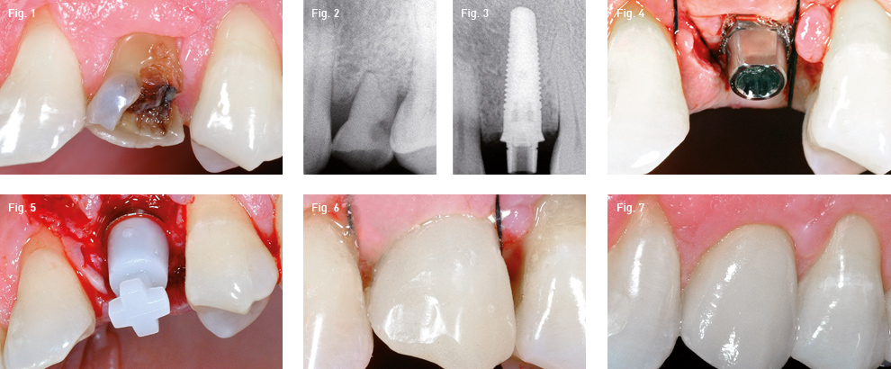
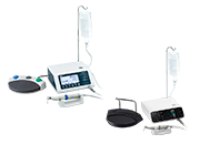
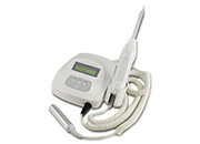

NumeriUno, 7: 17-18, 2010

According to the applicable rules and regulations, I declare under my own responsibilities to be a dentist and therefore I am authorized to read the contents of this web site.
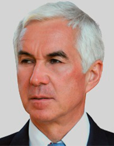
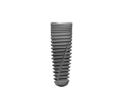
Master Degree in Medicine and Surgery at the University of Padova, specialization in Surgery. 3 years course in Orthodontics, dr Cozzani and dr Giannelli, Boston University. Update in Periodontics, Detroit University, prof Caffesse; update in Prosthesis, UCLA, prof Contino; course in Periodontics, dr Carnevale Active member AIOP. Private practice in Montagnana, Padova, particularly focused on surgery, implantology and implant prosthodontics.