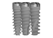
A 32 year old patient arrives at the dental office with a fractured element in position 15 and without the possibility of preservative recovering of the element. After having opted for a post-extraction procedure the patient is included in a socket preservation protocol and premature implant insertion since the patient did not have sufficient apical bone to anchor the implant so as to ensure the primary stability. After having performed the extraction in a most non-traumatic way possible, we proceed to the depithelization of the sulci with a diamond drill and to the filling of the socket with material (Bio-Oss® Collagen, Geistlich; Switzerland), which is compacted without using excessive force. The purpose to minimize the patient’s morbidity, a graft with collagen is used (Mucograft® Seal, Geistlich; Switzerland), which is sutured with simple stitches using a monofilament and nonresorbable suture.
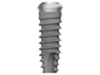
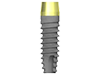
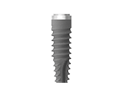
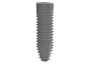
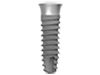
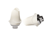
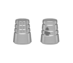
A week later we observe a good socket healing with integration of the graft. After two weeks the sutures are removed and a new epithelization at the edge of the socket is valued. After 4 weeks the total socket filling with a complete epithelization is observed. After 6 weeks from the frontal point of view it can be observed that the soft tissues have completed their maturation. After 8 weeks the implant insertion is planned, a CBCT is performed, in which sufficient bone availability is observed both in height as well as in thickness. After lifting a flap with total thickness, the maintenance of the bone structure in height and thickness after the preparation of the implant can be observed. A Premium TG Sweden & Martina implant of 3.8 x 11.5 mm with sufficient primary stability for a single surgical phase protocol and position a healing abutment is inserted. After one week the sutures are removed and after 8 weeks the impression is performed. From the X-ray image a correct osseointegration with maintenance of the interproximal bone level is valued. Finally, a tightened prosthetic structure in metal-ceramic (CAD-CAM) is manufactured. At the 6 months follow-up the partial filling of the papillae is valued.