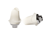
Patient B.L. of 83 years, male, past medical history with nothing to report except for pulmonary emphysema associated with fibrosis. The patient is a bearer of a partial mobile prosthesis with hooks to both jaw arches and of an endosseous tightened implant in position 1.4 inserted 8 years before, not subject to loading. The patient’s request was to have permanent teeth in the upper arch and a removable aesthetic prosthesis in the lower arch. When the appropriate evaluations were made, a treatment plan was suggested which included, respectively, a upper Toronto Bridge type prothesis supported by 8 posts, wrapped in titanium-composite, and a lower Valplast prosthesis with retention elements on the natural teeth. Under appropriate pharmacologic covering, at the time of the surgery we proceeded to the upper arch total cleaning, including the removal of the implant present in quadrant I and to the insertion of the planned fixtures. The distal ones with an adequate inclination for the purposes to extend the prosthetic use in the distal sense were positioned bilaterally, modifying the “tilted implants” concept, in this case limited by the vertical bones reduced diameters in correspondence to the bilateral sinus cavities (fact due to pneumatization of the same, associated with horizontal crestal atrophy). Where a discrepancy beyond one millimetre between the diameter of the used implant and that of the surgical extraction socket was encountered, the filling of the gap with an auto-heterologous graft was provided. The same areas were then recovered with a collagen membrane. After four months, during which the patient benefited from satisfying functional and aesthetic conditions thanks to the upper total prosthesis appropriately packaged, then we proceeded to the uncovering of the connective area of the implants, positioning appropriate healing abutments for the tissue conditioning. After two weeks, we proceeded to the impression taking with transfers tightened and to that of the other prosthetic parameters. The following sessions the insertion of the P.A.D. components on each single implant were included, benefiting from the aid of a guide key, verification and fitting of a titanium overstructure with an occlusal plate in composite, evaluating passivation, macroscopic level of fitting and the inter-maxillary congruity with an already fitted antagonist prosthesis. So finally we reached the set up and delivery session, where the prosthetic canals are sealed with teflon and composite.